 Suche
Suche
 Mein Konto
Porcine acute diarrhea syndrome coronavirus promotes replication by triggering autophagy

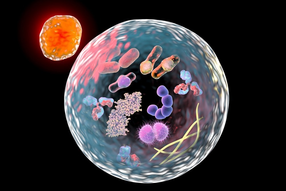
A recent iScience study finds that the porcine acute diarrhea syndrome coronavirus (SADS-CoV) promotes autophagy to maintain its replication in host cells. More specifically, the virus downregulates the AKT/mammalian target of rapamycin (mTOR) signaling pathway to trigger autophagy. Study: Acute porcine diarrhea syndrome coronavirus induces autophagy to promote its replication via the Akt/mTOR pathway. Image source: Kateryna Kon / Shutterstock.com Background SADS-CoV is an enveloped, single-stranded, positive-sense ribonucleic acid virus (RNA virus) that belongs to the Coronaviridae family. Other highly pathogenic members of the same virus family include severe acute respiratory syndrome coronavirus (SARS-CoV), Middle East respiratory syndrome coronavirus (MERS-CoV), and more recently...

Porcine acute diarrhea syndrome coronavirus promotes replication by triggering autophagy
A recent one iScience Study finds that porcine acute diarrhea syndrome coronavirus (SADS-CoV) promotes autophagy to maintain its replication in host cells. More specifically, the virus downregulates the AKT/mammalian target of rapamycin (mTOR) signaling pathway to trigger autophagy.
background
SADS-CoV is an enveloped, single-stranded, positive-sense ribonucleic acid virus (RNA virus) that belongs to the Coronaviridae family. Other highly pathogenic members of the same virus family include severe acute respiratory syndrome coronavirus (SARS-CoV), Middle East respiratory syndrome coronavirus (MERS-CoV), and more recently SARS-CoV-2.
SADS-CoV is a zoonotic coronavirus originating in bats that was recently discovered in 2017. The virus is potentially transmissible across species and can infect a range of cells derived from pigs, rats, monkeys and humans. This highlights the need to understand host-pathogen interactions to identify potential antiviral therapeutics.
Autophagy is an important host defense mechanism against invading viruses. This process helps destroy and eliminate viral components via the lysosomal degradation pathway. However, some viruses, such as Zika virus, human papillomavirus (HPV), and herpes simplex virus type 2, have been found to block host autophagy to promote replication and survival.
In the current study, scientists are investigating the connection between SADS-CoV infection and autophagy regulation.
Impact of SADS-CoV infection on autophagy
Monkey and pig-derived cells were infected with SADS-CoV-2 and subjected to autophagy analysis at different time points. Modulation of autophagy was assessed by estimating the expression of a vital autophagosome marker LC3-II.
SADS-CoV infection was found to induce expression of LC3-II at all time points tested postinfection. The highest expression was observed after 24 and 36 hours, depending on the cell type. Furthermore, microscopic analysis confirmed the accumulation of autophagosomes in response to SADS-CoV infection.
To determine whether a replication-incompetent virus can trigger autophagy, SADS-CoV was first inactivated by ultraviolet (UV) radiation and then used to infect cells. This experiment revealed that SADS-CoV needs to maintain its replication in host cells to stimulate the autophagy process.
The influence of autophagy on SADS-CoV replication was assessed using rapamycin and 3-methylamine, an established inducer and inhibitor of autophagy, respectively. While rapamycin was found to induce both autophagy and virus replication in host cells, an opposite effect was observed in cells treated with 3-methylademine.
These results suggest that SADS-CoV induces autophagy to facilitate its replication within host cells during infection.
Mechanism of SADS-CoV-induced autophagy
Autophagy is characterized by the formation of autophagosomes and the subsequent fusion of autophagosomes with lysosomes to degrade viral components. While some viruses induce fusion of autophagosomes with endosomes to survive, others prevent fusion between autophagosomes and lysosomes and subsequently inhibit autophagic flux.
A series of experiments to determine the mechanistic details of SADS-CoV-induced autophagy revealed that the virus induces complete autophagic flux to promote its replication. Inhibition of autophagosome-lysosome fusion was found to disrupt virus replication.
Further analysis revealed that SADS-CoV induces autophagy via the ATG5-dependent pathway. ATG5 is a protein required for the formation of autophagosomes. At the molecular level, SADS-CoV inhibited the AKT/mTOR signaling pathway to promote autophagy and maintain replication.
The mTOR signaling pathway plays a crucial role in the initiation of autophagy. AKT is a serine/threonine kinase that functions as an upstream signaling component of the mTOR signaling pathway to regulate autophagy.
Impact of autophagy inhibition on SADS-CoV replication
Proteomic analysis of SADS-CoV-infected cells was performed to identify potential antiviral targets. This led to the identification of eight differentially expressed proteins associated with the PI3K/AKT signaling pathway. Of these proteins, only integrin α3 (ITGA3) showed antiviral effects against SADS-CoV replication.
ITGA3 is a cell membrane adhesion protein closely related to autophagy. Overexpression of ITGA3 in SADS-CoV-infected cells resulted in downregulation of autophagy and viral replication and upregulation of AKT and mTOR activities. In contrast, opposite events were observed following suppression of ITGA3 in the virus-infected cell.
These results suggest that ITGA3 prevents SADS-CoV replication by inhibiting autophagy via the AKT/mTOR pathway.
Study Importance
The current study describes a novel mechanism of SADS-CV-induced autophagy that is required for virus replication and survival in host cells. Additionally, the study identifies ITGA3 as a potential antiviral molecule that can prevent virus replication by inhibiting autophagy.
Reference:
- Zeng, S., Zhao, Y., Peng, O., et al. (2022). Das Coronavirus mit akutem Schweinedurchfall-Syndrom induziert Autophagie, um seine Replikation über den Akt/mTOR-Weg zu fördern. iScience. doi: 10.1016/j.isci.2022.105394. https://www.sciencedirect.com/science/article/pii/S2589004222016662