 Suche
Suche
 Mein Konto
Congenital heart defects in children
Congenital heart defects in children
Total anomalous pulmonary venous return (TAPVR)
Total anomalous pulmonary venous return
Total anomalous pulmonary venous return
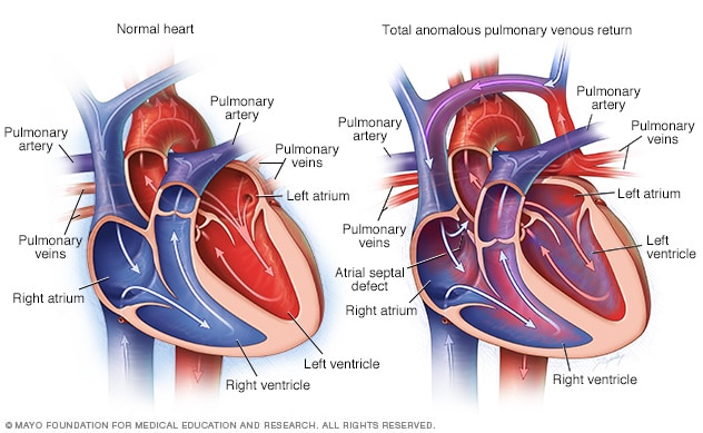
In total pulmonary venous return (TAPVR), the pulmonary veins mistakenly drain blood into the upper right chamber of the heart (atrium). In a normal heart (shown at left), oxygen-rich blood flows from the pulmonary veins into the upper left chamber (left atrium). In a type of TAPVR called supracardiac TAPVR, oxygen-rich blood flows from the pulmonary veins into the false chamber on the other side (right atrium). This causes the blood to mix with oxygen-poor blood.
Total pulmonary venous reflux (TAPVR) is a rare heart defect that is present at birth (congenital heart defect). It is sometimes called total anomalous pulmonary venous connection (TAPVC).
In this heart defect, the pulmonary blood vessels (pulmonary veins) are attached to the wrong place in the heart.
Normally, oxygen-rich blood travels from the lungs to the upper left chamber of the heart (left atrium) and then flows throughout the body. In TAPVR, blood is sent through an abnormal connection of veins instead through the upper right chamber of the heart (right atrium), where it mixes with deoxygenated blood. As a result, the blood flowing to the body does not have enough oxygen.
The specific type of TAPVR depends on where the veins connect. Most children born with TAPVR have no family history of congenital heart disease.
Symptoms
A baby with TAPVR may have difficulty breathing and appear blue (cyanotic) due to a lack of oxygen in the blood flowing to the body or because blood flow through the pulmonary veins is reduced or blocked.
A doctor may notice signs and symptoms of TAPVR shortly after birth. But some children don't have symptoms until later.
diagnosis
Your child's doctor will perform a physical exam and listen to your child's heart with a stethoscope to check for a heart murmur.
An echocardiogram is generally used to diagnose total anomalous pulmonary venous return. This test uses sound waves to create images of your child's moving heart. An echocardiogram can show pulmonary veins, holes in the heart, and the size of the heart chambers. It can also measure the speed of blood flow.
Other tests such as an electrocardiogram (EKG or EKG), chest x-ray, or computed tomography (CT) scan may be done if more information is needed.
Treatment
Surgery is generally required when a child is a baby. The timing of surgery depends on whether there is a blockage or not. To repair this congenital heart defect, surgeons connect the pulmonary veins to the left atrium and close the hole between the atria.
A person with total anomalous pulmonary venous return needs regular medical examinations with cardiologists trained in congenital heart disease to monitor infections, blockages, or heart rhythm problems.
Sources:
- Totale Lungenvenenverbindungsanomalie (TAPVC). American Heart Association. https://www.heart.org/en/health-topics/congenital-heart-defects/about-congenital-heart-defects/total-anomalous-pulmonary-venous-connection-tapvc#.Wc1Wh9jrvcs. Abgerufen am 12. April 2021.
- Dateien MD, et al. Total anomale Lungenvenenverbindung: Präoperative Anatomie, Physiologie, Bildgebung und interventionelles Management der postoperativen Lungenvenenobstruktion. Seminare in kardiothorakaler und vaskulärer Anästhesie. 2017; doi:10.1177/1089253216672442.
- Soriano BD, et al. Totale Anomalie der pulmonalvenösen Verbindung. https://www.uptodate.com/contents/search. Abgerufen am 12. April 2021.
- Fakten über den totalen anomalen pulmonalvenösen Rückfluss oder TAPVR. Zentren für die Kontrolle und Prävention von Krankheiten. https://www.cdc.gov/ncbddd/heartdefects/tapvr.html. Abgerufen am 12. April 2021.
- Paladini D, et al. Pränatale Diagnose einer totalen und partiellen Lungenvenenverbindungsanomalie: Multizentrische Kohortenstudie und Metaanalyse. Ultraschall in Geburtshilfe & Gynäkologie. 2018; doi:10.1002/uog.18907.