 Suche
Suche
 Mein Konto
Peripheral nerve tumors
Peripheral nerve tumors
overview
Schwannoma in the leg
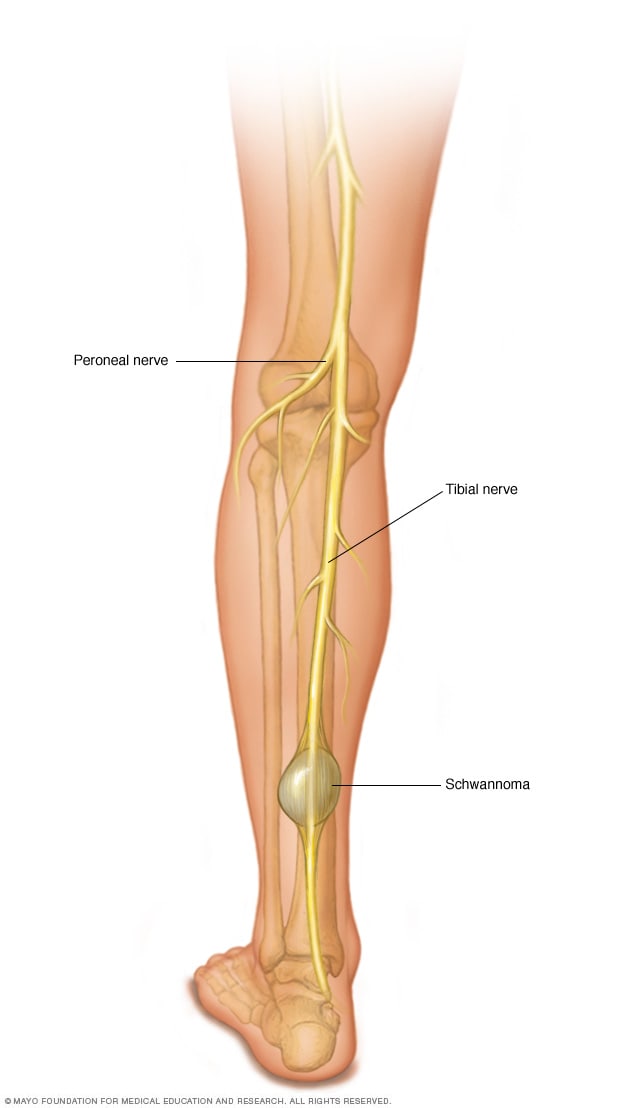
Schwannoma in the leg
Benign tumors can occur in nerves, muscles and bones. This illustration shows a schwannoma of the tibial nerve in the leg.
Dumbbell tumor

Dumbbell tumor
A more complex nerve sheath tumor may take the shape of a dumbbell. This type of tumor occurs in the spine and pelvis and is intertwined with important nerves.
Acoustic neuroma (vestibular schwannoma)
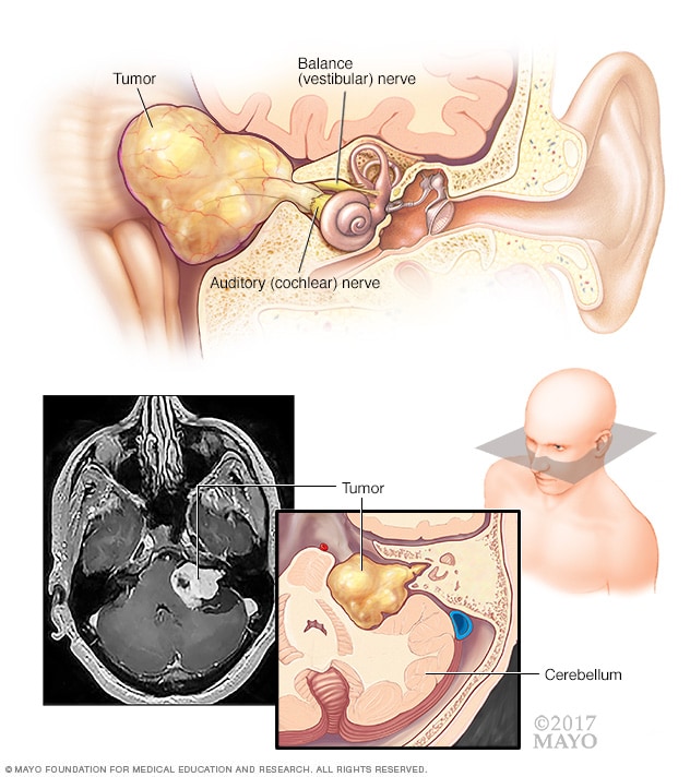
Acoustic neuroma (vestibular schwannoma)
An acoustic neuroma (vestibular schwannoma) is a benign tumor that develops on the balance and auditory nerves that run from your inner ear to your brain. These nerves are intertwined to form the vestibulocochlear nerve (eighth cranial nerve). The pressure from the tumor on the nerve can cause hearing loss and imbalance.
Peripheral nerve tumors are growths in or near the strands of tissue (nerves) that transmit signals from your brain to the rest of your body. These nerves control your muscles to allow you to walk, blink, swallow, pick up things, and perform other activities.
Peripheral nerve tumors can occur anywhere in the body. Most of them are not cancerous (malignant), but can cause pain, nerve damage, and loss of function in the affected area.
Treatment for peripheral nerve tumors usually involves surgery to remove the tumor. Sometimes the tumor cannot be removed without damaging nearby healthy tissue and nerves. In these cases, other treatments may be recommended.
Different types of peripheral nerve tumors occur. These tumors attack the nerves by growing within them (intraneural tumors) or pressing against them (extraneural tumors).
Types
-
Acoustic neuroma
-
Benign peripheral nerve tumor
-
Desmoid tumors
-
Malignant peripheral nerve sheath tumors
-
Neurofibroma
-
Neurofibromatosis
-
Schwannoma
Symptoms
The symptoms and signs of a peripheral nerve tumor develop from direct effects on the main nerve or from pressure from the tumor on adjacent nerves, blood vessels, or tissues. As the tumor grows, it may be more likely to cause signs and symptoms, although tumor size does not always determine the impact.
Signs and symptoms of peripheral nerve tumors vary depending on the location of the tumors and the tissues affected. They include:
- Schwellung oder ein Knoten unter der Haut
- Schmerzen, Kribbeln oder Taubheit
- Schwäche oder Funktionsverlust im betroffenen Bereich
- Schwindel oder Gleichgewichtsverlust
When to go to the doctor?
See your doctor if you have any of the symptoms listed, especially if you have a lump that is growing quickly.
Causes
Nervous system
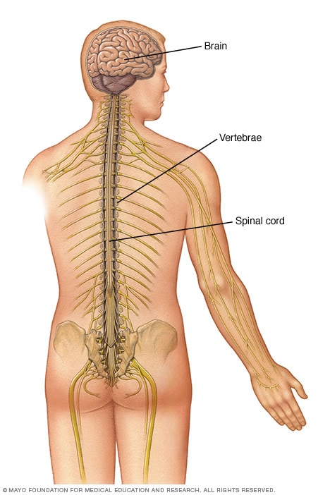
Nervous system
The spinal cord is housed in a hollow chamber within the vertebrae (spinal canal). It extends from the base of the skull to the lower back.
It is not clear why most peripheral nerve tumors develop. Some are associated with well-known inherited diseases such as neurofibromatosis (types 1 and 2) and schwannomatosis. Others can be caused by a faulty gene or triggered by injury or surgery.
Risk factors
Peripheral nerve tumors are more common in people with:
- Neurofibromatose (Typ 1 und 2) und Schwannomatose. Bei diesen Erkrankungen entwickeln sich Tumore an oder in der Nähe der Nerven im ganzen Körper. Diese häufig multiplen Tumoren können je nach Lokalisation zu unterschiedlichen Symptomen und Zeichen führen. Diese Tumoren sind in der Regel gutartig.
- Eine Geschichte der Strahlentherapie. Wenn Sie einer Strahlung ausgesetzt waren, haben Sie Jahre später ein erhöhtes Risiko für periphere Nerventumore.
Complications
Both benign and malignant peripheral nerve tumors can compress nerves, leading to complications, some of which may be permanent:
- Taubheit und Schwäche im betroffenen Bereich
- Funktionsverlust im betroffenen Bereich
- Schwierigkeiten mit dem Gleichgewicht
- Schmerzen
Treatment of peripheral nerve tumors
Sources:
- Gilchrist JM, et al. Tumore der peripheren Nerven. http://www.uptodate.com/home. Abgerufen am 28. Oktober 2016.
- Montano N. et al. Tumoren des peripheren Nervensystems: Analyse prognostischer Faktoren in einer Serie mit Langzeit-Follow-up und Literaturrecherche. Zeitschrift für Neurochirurgie. 2016;125:363.
- Riggin ER. Allscripts EPSi. Mayo Clinic, Rochester, Minnesota, 28. Okt. 2016.
- Überblick über Erkrankungen des peripheren Nervensystems. Merck Manual Professional-Version. https://www.merckmanuals.com/professional/neurological-disorders/peripheral-nervous-system-and-motor-unit-disorders/overview-of-peripheral-nervous-system-disorders. Abgerufen am 1. März 2017.
- Ahlawat S. et al. Magnetresonanzneurographie von peripheren Nerventumoren und tumorähnlichen Zuständen. Neuroimaging Clinics of North America. 2014;24:171.
- Klein CJ, et al. Genomanalysen zeigen häufige TRAF7-Mutationen bei intraneuralen Perineuriomen. Annalen der Neurologie. 2017;81:316.
- Amrami KK, et al. Einleitung: Bildgebung peripherer Nerven. Neurochirurgischer Schwerpunkt. 2015;39:E1. http://thejns.org/doi/pdf/10.3171/2015.6.FOCUS15314. Abgerufen am 8. April 2017.
- Spinner RJ, et al. Der vereinende artikuläre (synoviale) Ursprung der intraneuralen Ganglien: Evolution-Offenbarung-Revolution. Neurochirurgie. 2009;65:A115.
- Capek S. et al. Perineurale Ausbreitung von malignen Erkrankungen des Beckens bis zum lumbosakralen Plexus und darüber hinaus; Klinische und bildgebende Muster. Neurochirurgischer Schwerpunkt. 2015;39:E14.
- Carlson ML, et al. Schwannome des Gesichtsnervs: Überprüfung von 80 Fällen über 25 Jahre . Verfahren der Mayo-Klinik. 2016;91:1563.
- Ducatman BS, et al. Bösartige periphere Nervenscheidentumoren: Eine klinisch-pathologische Studie von 120 Fällen. Krebs. 1986;57:2006.
- Prasad, N., et al. Klinische Anatomie als Wegbereiter für Lösungen: Ein wichtiges Paradigma für die translationale Forschung. Klinische Anatomie. 2016;29:978.
- Broski SM, et al. Evaluation von 18-F-FDG-PET und MRT zur Differenzierung gutartiger und bösartiger Tumoren der peripheren Nervenscheide. Skelettradiologie. 2016;45:1097.
- Babovic-Vuksanovic D, et al. Multiple orbitale Neurofibrome, schmerzhafte periphere Nerventumoren, markantes Gesicht und marfanoider Habitus: Ein neues Syndrom. Europäische Zeitschrift für Humangenetik. 2012;20:618.
- Strahlenschädigung des Gehirns. International RadioSurgery Association. http://www.irsa.org/radiation_injury.html. Abgerufen am 11. April 2017.
- Papst TL. Weichteiltumoren. In: Muskuloskelettale Bildgebung. Philadelphia, Pennsylvania: Saunders Elsevier; 2015. https://www.clinicalkey.com. Abgerufen am 10. April 2017.
- Goldblum JR, et al., Hrsg. Gutartige Tumoren der peripheren Nerven. In: Weichteiltumoren von Enzinger und Weiss. 6. Aufl. Philadelphia, Pennsylvania: Saunders Elsevier; 2014. https://www.clinicalkey.com. Abgerufen am 10. April 2017.
- Koht A. et al. Neuromonitoring in Chirurgie und Anästhesie. http://www.uptodate.com/home. Abgerufen am 11. April 2017.
- Wise SC, et al. Chirurgische Bergung eines rezidivierenden Vestibularisschwannoms nach vorheriger stereotaktischer Radiochirurgie. Laryngoskop. 2016;126:2580.
- Stucky CCH, et al. Maligne periphere Nervenscheidentumoren (MPNST): Die Erfahrung der Mayo Clinic. Annalen der chirurgischen Onkologie. 2012;19:878.
- Spinner RJ (Gutachten). Mayo Clinic, Rochester, Minnesota, 28. Mai 2017.
- Überblick über die stereotaktische Radiochirurgie. International RadioSurgery Association. http://www.irsa.org/radiosurgery.html. Abgerufen am 11. April 2017.
-
Acoustic neuroma
-
Benign peripheral nerve tumor
-
Desmoid tumors
-
Malignant peripheral nerve sheath tumors
-
Neurofibroma
-
Neurofibromatosis
-
Schwannoma