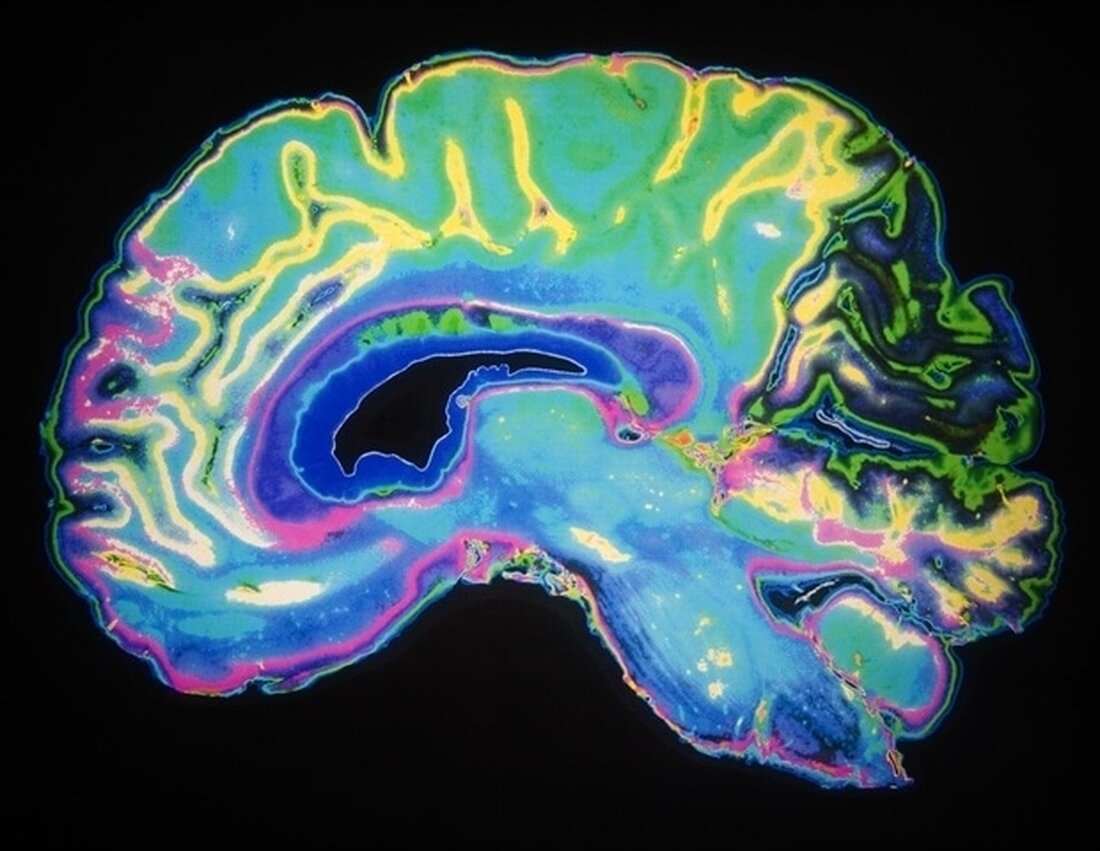
UH professor develops imaging system to improve view of cornea and fetal brain
Under the leadership of Kirill Larin, high-resolution optical coherence tomography (OCT) is experiencing a major upswing. Over the past 20 years, the UH biomedical engineering professor has advanced development from a device that examines only the retina to one that can measure an incredibly wide variety of internal organs, from the fetal heart to the neural tube. OCT does not require biopsies; No invasive measures are taken. Instead, the imaging technique uses light waves to capture cross-sectional images and provide 3D images. And while Larin has been pushing the boundaries of OCT's capabilities for more than two decades, he's just getting started. …

UH professor develops imaging system to improve view of cornea and fetal brain
Under the leadership of Kirill Larin, high-resolution optical coherence tomography (OCT) is experiencing a major upswing. Over the past 20 years, the UH biomedical engineering professor has advanced development from a device that examines only the retina to one that can measure an incredibly wide variety of internal organs, from the fetal heart to the neural tube.
OCT does not require biopsies; No invasive measures are taken. Instead, the imaging technique uses light waves to capture cross-sectional images and provide 3D images.
And while Larin has been pushing the boundaries of OCT's capabilities for more than two decades, he's just getting started.
Over $5 million in new investment in his work led him to develop OCT to look inside the fetal brain and assess how it is affected by maternal alcohol consumption and smoking. He is also bringing the technology into clinical settings to diagnose and prevent eye diseases.
The fetal brain – prevents and reverses the effects of alcohol and nicotine
Although the cause of congenital brain growth abnormalities is complex, prenatal alcohol/ethanol and nicotine exposure (PEE/PNE) are known as factors leading to such defects. To date, there has been a lack of sensitive, high-resolution tools to visualize dynamic changes in fetal physiology.
With a $3.2 million grant from the Eunice Kennedy Shriver National Institute of Child Health & Human Development, Larin and his team will develop a new sensitive, high-resolution imaging platform for in utero imaging of the fetal brain that will fill a significant gap in the developmental understanding of the emergence of brain growth deficits due to PEE/PNE.
Larin previously reported that prenatal ethanol exposure results in a rapid and sustained decrease in blood flow through the middle cerebral artery and pial perineural vascular plexus, which supplies blood to the fetal brain. Now he will explore this even further.
"We will develop a new imaging platform that combines the complementary advantages of OCT and two-photon light sheet microscopy (2pLSM) for in utero imaging of the fetal brain. The tools will allow us - for the first time - to go dynamic." , time-resolved assessment of capillary permeability and monocyte progenitor invasion,” said Larin.
“These studies will also enable us to begin evaluating the effectiveness of new pharmacological intervention strategies aimed at preventing or reversing the effects of alcohol, ethanol and nicotine exposure on fetuses.”
Insights into the cornea
With a $2.9 million grant from the National Eye Institute, Larin will take OCT in a further direction and develop a version of it that can be easily integrated into a doctor's practice.
"We propose a novel method for non-contact assessment of the elastic properties of the cornea. Such a technology, called heart-beat optical coherence elastography (hbOCE), could revolutionize methods for routine corneal examination, provide additional mechanical information and justify rapid clinical adaptation." " said Larin. "We will perform high-speed volumetric phase-sensitive OCT scans of the cornea during multiple phases of the heartbeat to measure corneal deformations and therefore biomechanics."
Accurate measurement of corneal biomechanics with high spatial resolution would not only influence the clinical interpretation of diagnostic tests, for example by measuring intraocular pressure or assessing the effect of drug therapies, but also predict the development of posterior eye diseases such as glaucoma. Currently, there is no reliable method to quantitatively measure corneal elasticity in vivo and with high resolution.
Our studies will accelerate the transition of ocular elastography into the clinic, inform our selection and use of corneal surgical treatments, and help us understand the structural consequences of corneal disease and wound healing.”
Kirill Larin, UH professor of biomedical engineering
Larin calls his work “frontier technology” because he pushes the boundaries of OCT and provides greater insight into the human body.
Source:
.

 Suche
Suche
 Mein Konto
